

The correct answer:
Case 26
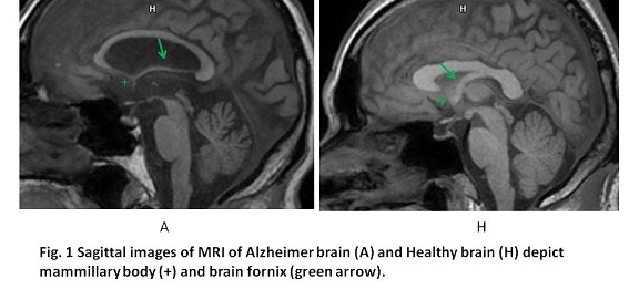
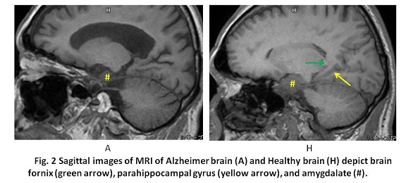
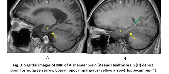
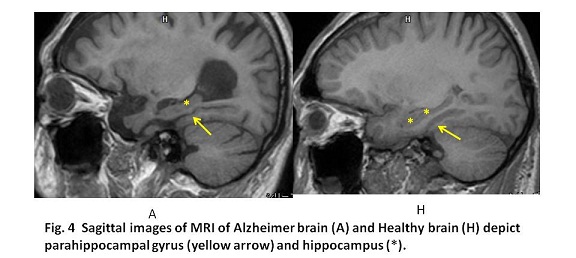
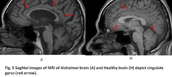
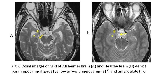
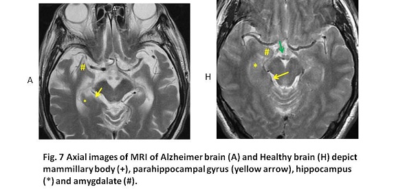
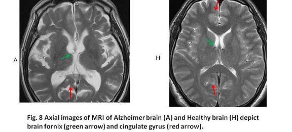
The recent research revealed that Alzheimer disease (AD) is triggered by deposition of tau protein, formation of neuro-fibrillary tangles and accumulation of β-amyloid (1). Further, carriers of the apolipoprotein E (ApoE)-ε4 genotype might be relevant with predisposition of AD (2). AD starts in a brain region known as lateral entorhinal cortex (LEC) in parahippocampal gyrus (1, 2). Namely, tau protein and β-amyloid precursor initially accumulate in the LEC and injures the neurons in the LEC. The LEC is considered to be a gateway to the hippocampus, which plays a key role in the consolidation of long-term memory (3). Thereafter, Alzheimer’s spreads from the LEC directly to the parietal cortex, posterior parietal association area (Area 7) via the perirhinal cortex. Area 7 involved in spatial orientation and navigation, and storage of long-term memory (1-5). Now that the onset site of AD was pinpointed, those changes might be observable using functional MRI, leading to detect early stage of AD (1, 3). Although functional MRI is unavailable in our hospital, the knowledge described above is useful to ensure imaging diagnosis of AD. MRI in our case with advanced AD showed marked volume loss of the limbic system including parahippocampal gyrus.
AD is the cause of 60% to 70% of cases of dementia (7, 8). The most common early symptom is difficulty in remembering recent events (short-term memory loss), implying disorder of entorhinal cortex. As AD advances, symptoms can include problems with language, disorientation (including easily getting lost), loss of motivation, not managing self care, and behavior issues (7, 8). As a screening index for AD, Mini-Mental Status Examination (MMSE) and Revised Hasegawa Dementia Scale (HDS-R) are available (9). In Japan, HDS-R is commonly used. Of the full mark of 30 points, the cutoff mark is 20 points for AD. Namely, the suspicious AD is 20 points or less and non-AD is 21 points or more (sensitivity 0.90, specificity 0.82). In our case, HDS-R was 14 points.
As objective numerical indicator for AD using MRI, voxel-based specific region analysis for AD (VSRAD) is available in Japan. First, voxels of gray matter were extracted from MRI image. Then, an objective brain is overlapped with the standard healthy brain. The voxel volume of parahippocampal cortex was used for comparison. As the standard brain, 80 healthy volunteers brains aged 54 to 86 years were used (10). Z score was calculated using the following formula: Mean voxels values of healthy hippocampal cortex – Mean voxels values of objective hippocampal cortex / Standard deviation among healthy hippocampal cortex values. Z score of 2 or greater means more than 2 fold of standard deviation, implying significant difference of P < 0.05 (10). Z score of VSRAD of our case was 3.83.
【Summary】
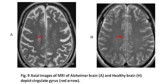
We presented two cranial MRIs for comparison between an Alzheimer brain and a healthy brain. Hasegawa criteria score for a patient with Alzheimer disease was 14 points and his voxel-based specific regional analysis for Alzheimer’s disease (VSRAD) was 3.83. The whole limbic system was atrophic in our case of AD, including parahippocampal gyrus ,lateral entorhinal cortex and posterior parietal association area.
【References】
1.Study Shows Where Alzheimer's Starts And How It Spreads Published: December 23, 2013. Released by Columbia University Medical Center
2.Khan UA, et al. Molecular drivers and cortical spread of lateral entorhinal cortex dysfunction in preclinical Alzheimer's disease. Nature Neuroscience. 2013 doi:10.1038/nn.3606.
3.Lopez, ME, et al. Alpha-Band Hypersynchronization in Progressive Mild Cognitive Impairment: A Magnetoencephalography Study. Journal of Neuroscience. 2014 34 (44):14551–14559. doi:10.1523/JNEUROSCI.0964-14.
4.Suthana, N, et al. "Memory Enhancement and Deep-Brain Stimulation of the Entorhinal Area". New England Journal of Medicine doi:10.1056/NEJMoa1107212.
5.Velayudhan L, et al. Entorhinal cortex thickness predicts cognitive decline in Alzheimer's disease. J Alzheimers Dis. 2013;33(3):755-66. doi: 10.3233/JAD-2012-121408.
6.Burns A, et al. "Alzheimer's disease".The BMJ. 2009; 338: b158. doi:10.1136/bmj.b158. PMID 19196745.(subscription required (help)).
7.World Health Organization."Dementia Fact sheet N°362". March 2015. Archived from the original on 18 March 2015. Retrieved13 January 2016.
8.National Institute on Aging "About Alzheimer's Disease: Symptoms". Retrieved 28 December 2011.
9.Kim KW, et al. Diagnostic accuracy of mini-mental status examination and revised hasegawa dementia scale for Alzheimer's disease. Dement Geriatr Cogn Disord. 2005;19:324-330. Epub 2005 Mar 22
10.Hirata Y, et al. Voxel-based morphometry to discriminate early Alzheimer's disease from controls. Neuroscience letters 382;3:269-274
2016.10.5
COPYRIGHT © SEICHOKAI YUJINKAI. ALL RIGHTS RESERVED.